DiATOME® Diamond Knives for CEMOVIS
(Cryo-Electron Microscopy Of Vitreous Sections)Available for sale in U.S.A. only
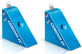 |
The CEMOVIS 35° knife and the CEMOVIS 25° knife are designed for sectioning frozen hydrated specimens. The 25° angle results in the least possible compression and the best structure preservation. Knife Specifications Knife angles: 25°, 35°Thickness range: 30-150nm Available size: 3mm |
Cleaning Procedure for DiATOME® CEMOVIS Diamond Knives Handling & Use of DiATOME® Diamond Knives (860KB PDF) |
|
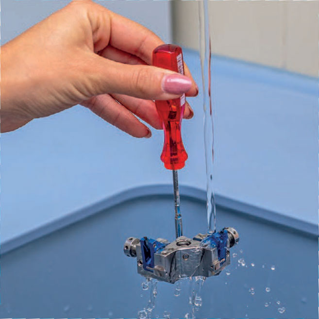
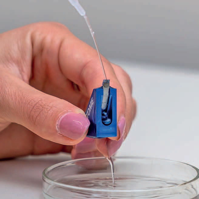
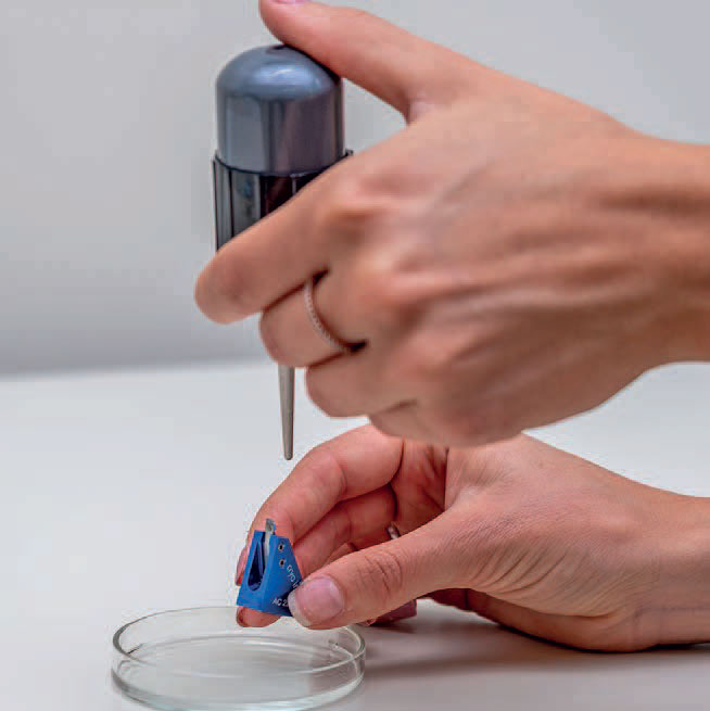
DiATOME® Cleaning Procedure for CEMOVIS Diamond KnivesThis procedure serves for the cleaning of our DiATOME® CEMOVIS knives. We are at your disposal for any further assistance you might require.
|
References
* Pierson, Jason, et al. "Improving the technique of vitreous cryo-sectioning for cryo-electron tomography: electrostatic charging for section attachment and implementation of an anti-contamination glove box." Journal of structural biology 169.2 (2010): 219-225.
* Han, H‐M., Benoît Zuber, and J. Dubochet. "Compression and crevasses in vitreous sections under different cutting conditions." Journal of microscopy 230.2 (2008): 167-171.
* Al-Amoudi, Ashraf, Daniel Studer, and Jacques Dubochet. "Cutting artefacts and cutting process in vitreous sections for cryo-electron microscopy." Journal of structural biology 150.1 (2005): 109-121.
* Michel, M., H. Gnägi, and M. Müller. "Diamonds are a cryosectioner's best friend." Journal of Microscopy 166.1 (1992): 43-56.
* Richter, Karsten. "Cutting artefacts on ultrathin cryosections of biological bulk specimens." Micron 25.4 (1994): 297-308.
* Zhang, P., et al. "Direct visualization of receptor arrays in frozen‐hydrated sections and plunge‐frozen specimens of E. coli engineered to overproduce the chemotaxis receptor Tsr." Journal of microscopy 216.1 (2004): 76-83.